Interesting Cases

What Factors Influence Persistent Neck Pain after Whiplash?

Whiplash injuries are a frequent cause of chronic neck and back pain and are common cases in the personal injury litigation arena.  Litigation of these cases is often predicated on science as much as it is predicated on the facts of the case.  The study below was recently published in Spine Journal, and will give some clarity about those factors that affect the development of chronic neck pain status post a rear-end motor vehicle accident.


Eulogio Pleguezuelos Cobo, MD; M. Engracia Pérez Mesquida; Elisabet Palomera Fanegas; Eva Moreno Atanasio; M. Beatriz Samitier Pastor; Cristina Perucho Pont; Carlos Matarrubia Prieto; Genoveva Reverón Gómez; Lluis Guirao Cano


Spine. 2010;35(9):E338-E343


Abstract and Introduction

Abstract

Study Design. Prospective longitudinal study.
Objective. To identify prognosis factors that allow us to identify patients with risk of developing chronic symptoms and disabilities after a whiplash injury.
Summary of Background Data. The prognosis factors for poor recovery in acute whiplash are not conclusive.
Methods. We included 557 patients who suffered whiplash injury after road traffic accident and visited the Department of Physical Medicine and Rehabilitation of Mataró Hospital (Spain) for medical evaluation and rehabilitation treatment. The variables were collected following a protocol designed for the study, and all patients were assessed through the Visual Analogue Scale (VAS) for the intensity of neck pain, the Goldberg Depression and Anxiety Scale and the Northwick Park Neck Pain Questionnaire (NPH) for cervical column functionality at initial evaluation and 6 months later.
Results. Factors related with VAS 6 months after the whiplash injury were women, age, number of days of cervical column immobilization, previous neck pain, self-employed workers, housewives, pensioners, students, presence of headache or dizziness, and VAS, Goldberg Depression and Anxiety scale, and NPH scores at initial evaluation. In multivaried analysis, it had been found that the variables that had influence on VAS 6 months after the whiplash injury were statistically significant for age, presence of dizziness, self-employed workers, and VAS and NPH scores at initial evaluation.
Conclusion. Our findings indicate that factors that allow us to identify patients at risk for poor recovery are age, dizziness, and initial evaluation of neck pain with VAS and cervical column functionality with NPH.

Introduction

The whiplash is a cervical column injury, which is caused when the neck is violently extended, generally produced in vehicle collisions. It was described first time by Crowe in 1928.[1] After the Quebec Task Force on whiplash-associated disorders (WAD) study, whiplash was defined as a set of symptoms that appear after an acceleration-deceleration mechanism of energy transfer to the neck. That energy transfer mechanism may be caused by road collisions between vehicles, but it could also occur in other circumstances (i.e., diving). The energy transferred results into bone or soft tissue injuries, which could invariably lead to a variety of clinical symptoms.[1]

The incidence is variable in different geographical areas.[1] In the United States, 3 cases per 1000 inhabitants are diagnosed of this disease per year,[2] in Norway, 2 cases per 1000 inhabitants per year,[3] in Australia, 1 case per 1000 inhabitants per year,[4] and in Quebec, 0.7 cases per 1000 inhabitants per year.[4]

The main characteristic of this syndrome is the absence of evidence of pathology findings as detected in different imaging techniques despite the traumatism intensity, symptoms, and clinical findings. This poor evidence carries along with important social and sanitary problems. Although we could say that whiplash is a benign pathology, we should also consider that this is a disease with an high impact on health public, because the huge number of incapacities produced are defrayed by the public coffers with an estimated expenditure to the tune of 10,000 million euros per year in Europe, whereas in the United States, the numbers could rise between 4.5 and 29 billion dollars.[5,6]

On the other hand, prognostic factors in whiplash injury can be either magnified or hidden because of the existence of court cases to obtain economic compensations. Cassidy et al[7] showed that there is a diminution in the incidence of whiplash, being up to 28% in those patients with a better prognostic, when compensation laws regarding accidents were modified. In the literature, there are no unified criteria to identify those patients with risk of developing chronic symptoms and disabilities in the whiplash injury. The goal of this prospective longitudinal study is to identify prognostic factors for poor recovery in whiplash injury after initial evaluation at Department of Physical Medicine and Rehabilitation (DPMR), considering pain as the main variable of the study.

Read more ...

43 year old with Headache and Double Vision-Medical legal implications

What are the medical legal implications of the following case?

A 43 year-old man presents to the emergency department (ED) complaining of a severe frontal headache that began suddenly and awakened him from sleep. The headache is associated with nausea, vomiting, and fevers. He also complains of new-onset diplopia (double vision) and photophobia (bright light bothers him), but denies any decrease in visual acuity. He denies experiencing any associated seizures, focal weaknesses, previous similar episodes, frequent headaches, or previous visual disturbances. He does not have any prior significant medical problems, and takes no medication. He drinks socially, does not smoke, and denies recreational drug use.

On physical examination, the patient is ill-appearing but alert and in no apparent distress. His vital signs reveal a temperature of 103.1°F (39.5°C), a blood pressure of 155/95 mm Hg, and a pulse of 110 bpm. The ocular examination demonstrates ptosis of the right eye (droopy eyelid), which is deviated inferolaterally and has a dilated and unreactive pupil. The visual field examination demonstrates bitemporal hemianopsia. Funduscopic examination shows normal venous pulsation and mild bilateral temporal disc pallor. The cranial nerves are otherwise without deficit. The neck is supple and without meningismus. Examination of the chest reveals mild bilateral gynecomastia, without nipple discharge. The lungs are clear to auscultation. Cardiac auscultation reveals a normal S1 and S2 and no murmurs, rubs, or gallops. The abdomen is soft and nontender, and no organomegaly is detected. Bilateral upper and lower extremity strength is 5/5, with normal deep tendon and plantar reflexes. The patient's sensation is intact to light touch and pinprick throughout, and the gait is normal.

Laboratory investigations reveal a hemoglobin concentration of 13 g/dL (130 g/L); a white blood cell (WBC) count of 16.0 × 10³/µL (16.0 × 10⁹/L), with 75% neutrophils; and a platelet count of 340 × 103/µL (340 × 109/L). The electrolyte, blood urea nitrogen (BUN), creatinine, and glucose examinations are all within normal limits. Cerebrospinal fluid (CSF) specimens show 420,000 red blood ceels (RBC)/μL, 20,000 WBC/μL, a normal glucose of 85 mg/dL (4.72 mmol/L), and an elevated protein concentration of 230 mg/dL (2.3 g/L). The CSF Gram stain is negative for bacteria. A computed tomography (CT) scan of the brain is performed, followed immediately by magnetic resonance imaging.

The radiologist misreads the films and the patient is given fever reducing medication and sent home by the emergency physician.  He is told to visit his family physician in a few days.  The following day, he is found unconscious and is returned to the hospital by ambulance.  He is admitted in critical condition and he is re-evaluated.  Re-evaluation included a re-examination of his imaging studies by other radiologists.

He is admitted to the ICU undergoes a surgical procedure, but he never regains consciousness, although his vital signs are stabilized.  He remained in a vegetative state.  The family sued the physicians and the hospital for malpractice.

Discussion

The noncontrast CT scan of the brain when re-reviewed showed a 2-cm sellar mass, with suprasellar extension. There was impingement on the optic chiasm and the hypothalamus, with upward displacement. There was increased density on the right side of the mass, which was suggestive of hemorrhage. The sagittal and coronal T1- and T2-weighted MRI scans demonstrated a large soft-tissue mass in the pituitary fossa, with areas of intermediate- and high-intensity signal suggestive of hemorrhage (see Figure 3). Coronal gadolinium-enhanced T1-weighted images revealed that the mass had a heterogeneous pattern of faint peripheral enhancement (Figure 4). There was evidence of mass effect on the right cavernous sinus, which was most evident in the coronal T1- and T2-weighted images. These findings are consistent with pituitary apoplexy as a result of hemorrhage with or without infarction, likely into a pituitary adenoma. Tests for evaluating the hormonal status of the patient revealed panhypopituitarism. Prior to the acute apoplectic episode, the patient had findings suggestive of central hypogonadism, probably as a component of his hypopituitarism caused by pituitary macroadenoma (diminished libido and bilateral gynecomastia). His neurologic finding (right-sided ptosis with a fixed and dilated pupil pointing downward and outward) was consistent with a right-sided 3rd nerve palsy caused by extension of hemorrhage into the right cavernous sinus.

Pituitary tumor apoplexy is defined as hemorrhage or infarction of a pituitary gland associated with the presence of a preexisting pituitary adenoma. It manifests as a sudden, severe headache, and it is sometimes associated with neurologic and hormonal dysfunction. The word "apoplexy" stems from a Greek term meaning to "have a stroke".[1] Neurologic symptoms and signs are secondary to displacement of the optic nerve and impingement of the 3rd, 4th, and 6th cranial nerves. Hormonal dysfunction results from destruction of the anterior pituitary gland.

Pituitary tumor apoplexy is a rare disorder with an annual incidence of about 1.2 per million.[2] Men are affected twice as often as women, and all age groups can be affected, with the majority of patients in the 5th or 6th decades of life.[3] It is estimated to occur in 1.5-27.7% of cases of pituitary adenoma.[4] Pituitary tumor apoplexy is only rarely associated with a healthy gland; however, approximately 50% of patients who present with pituitary tumor apoplexy are not diagnosed with a pituitary lesion prior to their presentation.[1] All types of pituitary tumors carry the same risk for apoplexy.

The most common symptom of pituitary tumor apoplexy is headache. Almost all patients describe a sudden, severe retro-orbital or bifrontal headache, which is associated with vomiting in two-thirds of cases.[4] The headache and vomiting result from the sudden increase in intrasellar pressure either caused by the hemorrhage or secondary to meningeal irritation from blood or tumor products that leak into the CSF. The increase in intrasellar pressure results in many of the symptoms and signs of pituitary tumor apoplexy.[5] Laterally, the increased pressure causes compression of the structures in the cavernous sinus, namely the 3rd, 4th, and 6th cranial nerves, with the 3rd being most commonly affected as a result of its vulnerable position (parallel to the lateral wall of the pituitary gland). The 6th cranial nerve is the least commonly involved because of its most lateral location within the sinus.

Ophthalmoplegia (caused by 3rd, 4th, and 6th nerve palsies or any combination thereof) is present in around 80% of patients presenting with pituitary tumor apoplexy.[4] Also located within the cavernous sinus is the trigeminal nerve; its involvement may cause facial pain or sensory loss. Carotid siphon compression may present as hemiplegia. Superiorly, the increased pressure compresses the optic chiasm, optic tract, or optic nerve, leading to decreased visual acuity or visual field defects (classically, bitemporal hemianopsia). Blood leaking into the subarachnoid space may result in chemical meningitis with fever, meningismus, and photophobia. Fever in patients with apoplexy may also be explained by alteration in thermal regulation caused by hypothalamic involvement by the hemorrhage or by adrenal insufficiency associated with hypopituitarism. Hemorrhage may extend into the brain parenchyma causing cortical irritation and provoking seizures.

The elevated intrasellar pressure also accounts for the endocrine abnormalities found in cases of pituitary tumor apoplexy. This pressure increase results in compression of the pituitary tissue, compromising its vascular supply and leading to hypopituitarism. Adrenal insufficiency is the most clinically significant result of hypopituitarism, contributing significantly to the mortality of patients with pituitary tumor apoplexy if not promptly recognized and treated. Although not common, patients with pituitary tumor apoplexy may have diabetes insipidus at presentation. The true etiology of diabetes insipidus in this setting is unknown, but it may result from the increased pressure on the pituitary infundibulum, which impedes the antidiuretic hormone from passing from the hypothalamus to the posterior lobe of the pituitary.

A precipitating factor is identified in 50% of cases of pituitary tumor apoplexy. Predisposing factors include dopamine agonist treatment, head trauma, pituitary irradiation, pregnancy, coronary artery bypass grafting, surgical operations, and anticoagulation. Endocrine stimulation tests are also associated with pituitary tumor apoplexy. It is postulated that hormones used in these tests may increase blood flow in pituitary adenomas, provoking bleeding in friable vessels. Pituitary tumor apoplexy following childbirth associated with significant postpartum hemorrhage in nontumorous glands is termed "Sheehan syndrome". The hypertrophy of the pituitary gland that occurs in normal pregnancy combined with the arterial spasm of the pituitary's blood supply (caused by bleeding and hypotension) both contribute to the development of Sheehan syndrome; however the clinical presentation of pituitary apoplexy in these cases is usually less dramatic, with a more gradual development of signs and symptoms of hypopituitarism.

The diagnosis of pituitary tumor apoplexy is best established by MRI; however, this is usually preceded by a rapid diagnostic CT scan to screen for intracranial hemorrhage. MRI is superior to CT scanning for evaluating the pituitary gland and possibly visualizing hemorrhage not seen by CT. In one study, the detection rate of pituitary tumor apoplexy by CT scanning was 21%, whereas the detection rate was 100% with MRI.[3]

Once recognized, effective treatment of pituitary tumor apoplexy requires prompt administration of high-dose corticosteroids. Steroids should be administered in supraphysiologic doses to not only replace endogenous hormone deficiency during a stressful condition, but also to take advantage of its anti-inflammatory effect by decreasing swelling on parasellar structures. The definitive treatment for pituitary tumor apoplexy is emergent surgical decompression. Transsphenoidal resection is the most common approach in this situation. In cases where there is significant extension of hemorrhage into the brain parenchyma beyond the diaphragma sella, an intracranial approach may be preferred. In a minority of cases, conservative medical therapy is an acceptable alternative; examples of this include patients who are poor surgical candidates and selected patients who present with isolated meningismus or ophthalmoplegia and show significant improvement with steroid administration. Medical management includes monitoring of endocrine, neurologic, and ophthalmologic function combined with hormone replacement.

With prompt recognition, timely surgery, and proper medical management, the majority of patients with pituitary tumor apoplexy improve.[6] Ophthalmoplegia is usually the first symptom to resolve. Less readily restored is the optic nerve defect resulting in decreased visual acuity and restricted visual fields. More than half of patients, however, will have permanent hormone deficiencies resulting from pituitary injury and will require hormone replacement. One study showed that maintenance steroid, thyroid hormone, and testosterone replacement was essential postoperatively in 82%, 89%, and 64% of patients, respectively.[7]

Following immediate administration of high dose corticosteroids, the patient in this case underwent an emergent transsphenoidal resection. An infarcted adenoma was identified, with extensive areas of hemorrhage and necrosis consistent with apoplexy. After surgery the swelling in the brain worsened and he remained in a coma. An endocrinology evaluation was completed, and the patient was confirmed to have hypopituitarism, but suffered the effects of significant intracranial bleeding.

The plaintiff's theory of the case was that a correct diagnosis would have resulted in immediate surgery before the patient had bled to a degree where he has lost brain function and consciousness.  The jury agreed.  The case was taken to court where a plaintiff verdict was returned.


References

  1. Verrees M, Arafah B, Selman WR. Pituitary tumor apoplexy: characteristics, treatment, and outcomes. Neurosurg Focus. 2004;16:E6.
  2. Nielsen EH, Lindholm J, Bjerre P, et al. Frequent occurrence of pituitary apoplexy in patients with non-functioning pituitary adenoma. Clin Endocrinol (Oxf). 2006;64:319-22.
  3. Randeva HS, Schoebel J, Byrne J, Esiri M, Adams CB, Wass JA. Classical pituitary apoplexy: clinical features, management and outcome. Clin Endocrinol (Oxf). 1999;51:181-8.
  4. Vaphiades MS. Pituitary Apoplexy. eMedicine from WebMD. Last Updated: August 6, 2009. Available at: http://emedicine.medscape.com/article/1198279-overview.
  5. Zayour DH, Selman WR, Arafah BM. Extreme elevation of intrasellar pressure in patients with pituitary tumor apoplexy: relation to pituitary function. J Clin Endocrinol Metab. 2004;89:5649-54.
  6. Dubuisson AS, Beckers A, Stevenaert A. Classical pituitary tumour apoplexy: clinical features, management and outcomes in a series of 24 patients. Clin Neurol Neurosurg. 2007;109:63-70.
  7. Veldhuis JD, Hammond JM. Endocrine function after spontaneous infarction of the human pituitary: report, review, and reappraisal. Endocr Rev. 1980;1:100-7.


De-escalating the Toxicologically Altered Aggressive Individual

Toxicologists are often faced with cases where the subject in question had altered mental status as a consequence of the effects of illicit substances.  In this setting, dealing with aggressive patients can make a big difference in outcome. Patient death or injury resulting from the use of restraint and seclusion is an increasing concern in the field and in prison. Excessive and inappropriate TASER use has also been associated with sudden death.  A well-known 1998 article[1] documented 142 restraint-related deaths nationwide over a decade, 40% of which were attributed to unintentional asphyxiation during restraint. Restraint not only poses a risk for patient harm but also is physically and emotionally traumatizing for staff involved in the incident. Stefan pointed out that "high restraint rates are now understood as evidence of treatment failure."[2] Since the Joint Commission began tracking sentinel events in 1996, it has reviewed the deaths of 20 patients who were physically restrained.[3] Since then, the Joint Commission has advocated standards based on prevention as an intervention and the use of restraint as a last resort only after the least restrictive measures are exhausted.

Most communities have a protocol to call for team assistance when a psychiatric patient begins to display aggression or when an ordinarily calm individual becomes agitated while on excitatory drugs such as methamphetamine, cocaine, or phencyclidine. Law enforcement often believe that there is power in numbers, which can be true in certain situations. However, the increased external stimuli of gathering more police officers can also have untoward effects on the patient. The show of force may contribute to the escalation of combative behaviors.

Evidence points to a direct correlation between a high level of anxiety or perceived powerlessness on the patient's part and ensuing aggression.[4] The underlying cause of the behavior should be readily identified and handled accordingly. For instance, patients can become angry as a result of hallucinations, external provocation, or physical discomfort.

The Third-Person Approach

Although restraint may be necessary in emergency situations for patient and first responder safety, physical confrontation can usually be averted if de-escalation techniques are implemented before the patient gets out of control. De-escalation using a third-person approach, if implemented judiciously and cautiously by first responders, can be very effective in managing patients in the early stages of anger and aggression.

The third-person approach is similar to hostage crisis negotiation, in which a third party is brought in to negotiate a solution. Usually, it is much easier for the third person to take a neutral stance and to allow space for the angry person to step down. Billikopf postulates that all other things being equal, an outside third party has a greater chance than an insider of successfully mediating and resolving a difference.[6] The third person is not an arbiter trying to decide right from wrong, but a nonjudgmental facilitator of communication.

A "third party" or "third person" is a trained crisis interventionalist who was not present at the start of the dispute or conflict. A person who was involved in the conflict may be perceived, from the patient's standpoint, as being part of the problem. The ideal third person is someone who knows the patient well and with whom the patient has a certain degree of rapport.

The value of a therapeutic relationship has been a known and established fact for many decades. Research suggests that ineffective interpersonal relationships and interactions are major factors in escalating the aggressive behavior of a volatile individual.[8,9] Irwin concludes that intolerable environments and ineffectual interactions are far more likely to influence behaviors than are psychiatric symptoms alone.[10]

Use of the Third Person in De-escalation

Whenever an outburst is anticipated, the audience should be removed immediately. If team assistance is called in accordance with institutional policy, it may be better for the team members to stay in the background, ready to provide support when needed, but allow a single, third person from the care team to approach the patient. This less-than-expected response, or "under-reaction," can promote de-escalation.[11] The Pennsylvania Patient Safety Authority also suggests shifting the method of intervention from "a show of force to a show of support."[12] A 3-month study on the use of least restrictive interventions found that patients commonly select "verbal warning or talking things through" as the most valuable tool of anger management.[13] In short, the men (and women) in blue need to put their guns away and back off.  Crisis intervention teams will have greater success at de-escalating any situation, and there will be much less risk of injury to the patient and to law enforcement.

The third person should maintain a calm and supportive demeanor and use therapeutic communication skills. Avoid arguing with the patient or getting into a power struggle, and listen with empathy; the Greek Stoic philosopher Epictetus said that we have 2 ears and 1 mouth, so that we can speak less and listen more. In addition, state everything in clear, simple language: As anger escalates, the patient's perceptual field becomes limited; he or she probably cannot understand complex reasoning or process what you are saying. Tell the patient that you want to help, but he or she needs to calm down first. It is appropriate to say something like, "I would like to help you, but I can't hear you if you are screaming and yelling." Do not react to verbal attacks from the patient. Be aware of your own feelings of countertransference.

Staff members who take on the role of third person should have proper training in various techniques of nonviolent crisis intervention. The third person must also practice safety precautions, such as standing beyond arm's reach of the patient, positioning himself or herself for easy escape, and avoiding displays of body language that may be viewed as provocative to the patient.

Sometimes patients act out because they feel threatened. Assure the patient that he or she is safe, then set firm but nonthreatening limits. Offer choices to gain the patient's cooperation, and present positive reinforcement first. Positive reinforcement does not have to be a material reward; it can be praise and encouragement, or earning a certain privilege. In Rosenheck and Neale's 6-month study of 40 Veterans Affairs Assertive Community Treatment Program teams, clients with violent behavior who were exposed to negative limit-setting interventions typically had poorer outcomes.[14]

First responders have an obligation to maintain the safety of the patient and others in the environment. If restraint is deemed necessary, it should be used only when all measures of de-escalation have failed. In reality, no rigid policy or clinical guideline can spell out each and every scenario when physical restraint is the lesser of 2 evils. Crisis intervention workers have to rely on their own clinical judgment to weigh the risks and benefits of the measures they are considering. When to initiate physical restraint is a situation that depends on circumstances.[15]

References:

1. Weiss EM. Deadly restraint: a Hartford Courant investigative report. Hartford Courant. 1998;October 11-15. Section A.1 Available at" http://courant.ctnow.com/projects/restraint/data.stm


2. Stefan S. Legal and regulatory aspects of seclusion and restraint in mental health settings. Special edition: Violence and Coercion in Mental Health Settings: Eliminating the Use of Seclusion and Restraint. NTAC Networks. 2002;Summmer/Fall


3. Massachusetts Coalition for the Prevention of Medical Errors. Principles and Best Practice Recommendations to Improve Patient Safety Related to Restraint and Seclusion Use. 2003. Available at: http://www.macoalition.org/documents/RestraintPrinBestPrac-Definitions-Final.pdf Accessed on December 24, 2009.


4. Johnson B, Martin M, Guha M, Montgomery P. The experience of thought-disordered individuals preceding an aggressive incident. J Psychiatr Ment Health Nurs. 1997;4:213-220.


5. Davidson SE. The management of violence in general psychiatry. Advances in Psychiatric Treatment. 2005;11:362-370.


6. Billikopf G. Chapter 4: Interpersonal negotiation skills. In: Party-Directed Mediation: Helping Others Resolve Differences. Modesto, California: Regents of the University of California; 2009:85.


7. Peplau HE. Interpersonal Relations in Nursing. A Conceptual Frame of Reference for Psychodynamic Nursing. London: Macmillan; 1998.


8. Breeze J, Repper J. Struggling for control: the care experiences of “difficult” patients in mental health services. J Adv Nurs. 1998;28:1301-1311.


9. Secker J, Benson A, Balfe E, Lipsedge M, Robinson S, Walker J. Understanding the social context of violent and aggressive incidents on an inpatient unit. J Psychiatr Ment Health Nurs. 2004;11:172-178.


10. Irwin A. The nurse's role in the management of aggression. J Psychiatr Ment Health Nurs 2006;13:309-318.


11. Kreisberg L. Constructive Conflicts. Lanham, Md: Rowman & Littlefield; 1998:181-222


12. Pennsylvania Patient Safety Authority. The PA-PSRS Patient Safety Advisory. 2005; 2:1-6. Available at: http://patientsafetyauthority.org/ADVISORIES/AdvisoryLibrary/2005/Mar2(1)/Documents/22.pdf Accessed December 24, 2009.


13. Morales E, Duphorne PL. Least restrictive measures: alternatives to four-points and seclusion. J Psychosoc Nurs Ment Health Serv. 1995:33:13-16.


14. Rosenheck RA, Neale MS. Therapeutic limit setting and six-month outcomes in a Veterans Affairs assertive community treatment program. Psychiatric Serv. 2004;55:139-144.


15. Mohr WK, Petti TA, Mohr BD. Adverse effects associated with physical restraint. Can J Psychiatry. 2003:48:330-337.

A Patient with Pneumonia and Severe Arthralgias and other Findings. Is Failure to DIagnose Medical Malpractice?

Sometimes diseases are so rare and case presentations so atypical that it is difficult to blame a physician for failure to diagnose, and this should not lead to a medical malpractice action, no matter what the field: emergency medicine, primary care, or medical specialty care.  Here is a case in point:

A 61-year-old man presents to the emergency department (ED) with fever, dyspnea and a productive cough for 1 week that has failed to respond to outpatient antibiotics (levofloxacin). He also complains of worsening arthralgias in both lower extremities, particularly in his knees and ankles, as well as a 10-lb (4.54-kg) weight loss over the preceding 2 months. He is a former smoker with an 80-pack-year history, but there is no other significant medical history. He denies any recent travel, sick contacts, or occupational exposure to asbestos or mineral dust.  His only medications include over-the-counter analgesics for joint pains, and he denies having any drug allergies.

Read more ...

EMTALA Violations, ER Overcrowding, and Litigation

The following excerpt on EMTALA (Emergency Medical Treatment and Active Labor Act) is taken from a paper written by Dr. Damon Dietrich and Dr. Michael Crapanzano. The paper is entitled, "Emergency Department Diversion and Overcrowding: A Public Health Crisis." The paper discusses EMTALA in the context of ED diversion (EDD) and ED overcrowding (EDO).

In the U.S. today with increasing ER patient burden EMTALA violations have increased. This has resulted in increased litigation as well.

EDO and EDD are two of the most critical public health issues facing our nation's healthcare system today.[3,4,5]

  • ED visits in 2003 rose to 114 million, up from 97 million in 1997.
  • While visits increased by 17% over six years, 1128 EDs closed between 1988 and 1998, thus resulting in a dramatic increase in patient volumes and waiting times.
  • US hospitals over the past ten years have closed more than 103,000 inpatient medical/surgical beds and 7800 ICU beds in an effort to control costs.
  • The majority of the nation's 4000 hospitals and EDs report operating at or above critical capacity.
  • In 2001, two out of every three hospitals reported diverting ambulances to other hospitals.
  • EDO is reported to be most severe in areas with larger populations, higher population growth and higher than average uninsured patient volumes.

EDs represent the most critical access path to the nation's health delivery system, as the "guaranteed access point for all who need care regardless of ability to pay."[2] EDO exists when the ED has more patients than bed capacity or is over-saturated; this is a warning sign of capacity constraints under normal conditions. The March 2003 General Accounting Office Report indicated that EDO has many negative implications with regard to quality of care including prolonged patient wait times and suffering for acute problems while other patients are "boarded" in the ED, higher physician and staff stress, less confidentiality when patients are evaluated in nontraditional locations such as a hallway or on the EMS stretcher and increased transport times for ambulance patients due to diversion.[5] Contrary to public misconception, the gridlock in the ED is not in the waiting room, but rather occurs in the hallways of the ED with admitted patients waiting for beds upstairs for hours to days! The purpose of EDD was an innovative solution for EDO intended to "divert" stable patients transported by ambulance away from the hospital, thereby allowing the scarce beds remaining in the ED to be used for critical or unstable patients. EDD has been defined by The Lewin Group.[2]


Read more ...

Missed Diagnosis of Pulmonary Emboli

Hypothetical Case Study:

A 39-year-old woman presents to the emergency department (ED) with a non-productive cough, non-pleuritic chest pain, and shortness of breath (dyspnea). She is a smoker currently using Estrogen for hormonal abnormalities, but has no other significant past medical history. Her physical examination is unremarkable except her pulse which is mildly elevated, including clear lung auscultation and a non-tender chest wall. Although you contemplate the diagnosis of pulmonary embolism, her well appearance and normal vital signs notwithstanding pulse including normal oxygen saturation argue for bronchitis-related symptoms. You consider and then order a D-dimer study to rule in or rule out a pulmonary embolus (clot in the pulmonary arteries).

Clinical Question

Can a subset of patients with sufficiently low risk for pulmonary embolism be identified who require no diagnostic testing or should all patients that have a clinical picture to some degree consistent with pulmonary emboli be fully evaluated?

Read more ...

Ruptured Brain Aneurysms and Subarachnoid Hemorrhage-a Comprehensive Review

A superb analysis of ruptured brain aneurysm and subarachnoid hemorrhage (SAH) was written by Edlow et al and was published in the Journal of Emergency Medicine, volume 34 (3), 2008. The article is reprinted and shared with you in its entirety below. References are include.

Summary:

Aneurysmal subarachnoid hemorrhage (SAH) is a serious cause of stroke that affects 30,000 patients in North America annually. Due to a wide spectrum of presentations, misdiagnosis of SAH has been reported to occur in a significant proportion of cases. Headache, the most common chief complaint, may be an isolated finding; the neurological examination may be normal and neck stiffness absent. Emergency physicians must decide which patients to evaluate beyond history and physical examination. This evaluation–computed tomography (CT) scanning and lumbar puncture (LP)–is straightforward, but each test has important limitations. CT sensitivity falls with time from onset of symptoms and is lower in mildly affected patients. Traumatic LP must be distinguished from true SAH. Cerebrospinal fluid analysis centers on measuring xanthochromia. Debate exists about the best method to measure it–visual inspection or spectrophotometry. An LP-first strategy is also discussed. If SAH is diagnosed, the priority shifts to specialist consultation and cerebrovascular imaging to define the offending vascular lesion. The sensitivity of CT and magnetic resonance angiography are approaching that of conventional catheter angiography. Emergency physicians must also address various management issues to treat or prevent early complications. Endovascular therapy is being increasingly used, and disposition to neurovascular centers that offer the full range of treatments leads to better patient outcomes. Emergency physicians must be expert in the diagnosis and initial stabilization of patients with SAH. Treatment in a hospital with both neurosurgical and endovascular capability is becoming the norm.

Read more ...

What happened to the SHAVING BLADE!

Background

Figure 1.
Figure 1.
(Click to enlarge)
Figure 2.
Figure 2.
(Click to enlarge)

A 41-year-old cognitively impaired man who lives in a home for the mentally impaired is presented to the emergency department (ED) by his caregivers. His caregivers at the home noticed that, after washing the patient, 1 of 2 shaving blades present in the morning was missing. Although they hadn't witnessed it, they suspected that the patient had ingested the missing shaving blade. The caregivers, however, haven't noticed anything abnormal with the patient; he does not have any shortness of breath, stridor, wheezing, hemoptysis, vomiting, or upper gastrointestinal (GI) bleeding. At no point have the caregivers noticed the patient coughing or choking; however, they are still worried about the possibility that he might have ingested or aspirated the blade.

At baseline, the patient has unintelligible speech and, therefore, cannot be directly interviewed. The patient has been living in a residence for the mentally impaired for the past 31 years, and he is dependent on his caregivers for all of the activities of daily living. Apart from his mental underdevelopment, the patient suffers from epilepsy and has been previously investigated for Raynaud's disease; in addition, the patient's history is significant for an ejection systolic murmur. The murmur was first detected by echocardiography, which revealed normal left ventricular function and a slightly thickened mitral valve, with minimal regurgitation. He takes multiple medications, including dipyridamole 100 mg TID, phenytoin 100 mg TID, doxepin 25 mg BID, nitrazepam 5 mg nocte, carbamazepine 20 mg BID, nifedipine 10 mg BID, nortriptyline 25 mg TID, and risperidone 1 mg nocte.

On physical examination, the patient is not in any distress. His oral temperature is 98.6°F (37.0°C). His pulse is regular at 82 bpm and his blood pressure is 140/90 mm Hg. His oxygen saturation is 99% while breathing room air. A visual inspection of the oropharynx does not reveal the presence of a foreign body, and there is no evidence of any recent trauma, bleeding, or swelling. There is normal range of motion in the neck, and no evidence of stridor or respiratory difficulty is noted. On chest auscultation, there is good air entry bilaterally, and normal vesicular breathing is heard. His heart sounds reveal a 2/6 ejection systolic murmur. His abdomen is soft and nontender. Additionally, no distention or rebound is noted on the abdominal examination.

Read more ...

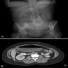
Woman with Sudden Onset Nausea and Vomiting: Could this be a case for a Toxicology Expert Witness?

Background
Figure 1.
Figure 1.
(Click to enlarge)

A 46-year-old woman presents to the emergency department (ED) with a history of worsening, constant right upper quadrant pain that radiates to her back and side. She has had nausea and has vomited twice in the past several hours. The intake triage nurse considers that she may have ingested something toxic, and turns immediately to her Emergency Physician who is also a well known toxicology expert consultant.  But he learns upon questioning that she underwent a laparoscopic cholecystectomy 2 weeks ago, without complications, and returned to her normal diet. She has not had any bowel movements or flatulence since the pain began. She denies having any fever, chills, or rigors. Her medical history is significant only for high blood pressure, high cholesterol levels, and gallbladder disease. She takes lisinopril, aspirin, multivitamins, and ginseng. She denies smoking or drinking alcohol.

On physical examination, the patient is awake, alert, and oriented. Her vital signs are within the normal range, with a heart rate of 84 bpm and a blood pressure of 124/76 mm Hg. She appears to be in mild distress. The cardiorespiratory examination yields normal findings, with clear lungs and a regular heart rhythm. Her abdomen is soft, but her bowel sounds are decreased, and she has marked tenderness in the right upper quadrant. The rest of her abdomen is minimally tender, with no evidence of guarding or rebound and no palpable masses. The other physical findings are normal.

The laboratory investigation reveals an elevated WBC count of 14.0 × 10⁹/L (14.0 × 10³/µL), with a left shift of 87% neutrophils. Her liver function tests, lipase level, and basic chemistry panel are unremarkable.

Contrast-enhanced computed tomography (CT) scans of the abdomen and pelvis are ordered. Figure 1A shows an anteroposterior (AP) scout image, and Figure 1B shows a selected axial section. The appearance of the xrays is that of bean-like substances.

The diagnosis is Cecal Volvulus.


Read more ...

Copyright © 2020 - www.emergencymedicineexpert.com & Dr. Barry Gustin